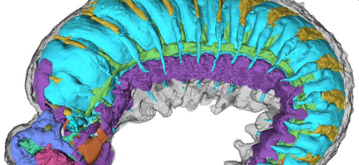
Researchers have uncovered the internal anatomy of a prehistoric creature the size of a poppy seed in extraordinary detail using powerful X-rays to scan a 520-million-year-old fossil. The findings, published in the journal Nature, reveal microscopic blood vessels and a nervous system, providing insight into one of the earliest ancestors of modern insects, spiders, and crabs.
X-rays reveal tiny half-billion-year-old creaturehttps://t.co/0iRkGlk2Lh
— William Rory Coker (@CokerRory) July 31, 2024
Lead researcher Dr. Martin Smith expressed his excitement about the discovery, noting that the fossil was preserved in its larval, or immature, stage when its body was still developing. He explained that studying these early stages is crucial to understanding how adult body shapes are formed, both through evolution and development. Dr. Smith remarked, “The chances of finding a fossilized larva are practically zero—or so I thought.”
Dr. Smith’s colleagues discovered the fossil in a pile of “prehistoric grit” while studying half-billion-year-old rock deposits in northern China, known for containing microscopic fossils. The team at Yunnan University spent years sifting through the material, eventually uncovering the exceptional specimen.
Dr. Smith realized the fossil’s significance after examining it under a microscope during a visit to China. He brought the specimen back to the UK for a closer examination using intense X-rays at Oxford’s Diamond Light Source facility. The team mounted the fossil on the head of a pin to scan it, revealing its internal secrets.
Dr. Smith was astonished by the structures preserved under the fossil’s skin. The researchers generated three-dimensional images of its miniature brain regions, digestive glands, a primitive circulatory system, and traces of the nerves supplying the larva’s simple legs and eyes. The segmented brain cavity provided insights into the ancestral features of modern insects, spiders, and crabs, which later evolved into specialized appendages like antennae, mouthparts, and eyes.
Study co-author Dr. Katherine Dobson from the University of Strathclyde praised the fossil’s preservation, achieved through natural fossilization. Dr. Smith suggested that high concentrations of phosphorus in the ocean where the larva lived and died might have contributed to its preservation. He explained, “The phosphorus seems to have flooded the tissues of our fossil,” essentially crystallizing its tiny body.
This discovery offers a rare glimpse into the early development stages of ancient arthropods, shedding light on the evolutionary processes that led to the diverse body structures seen in modern insects, spiders, and crabs. The study exemplifies the power of advanced imaging techniques in uncovering the hidden details of prehistoric life, providing valuable information about the history of life on Earth.
520-million-year-old worm fossil solves mystery of how modern insects, spiders and crabs evolved
https://t.co/E2AnIAIrAx— Pepe Muñoz (@PepeMunoz1) July 31, 2024
Major Points:
- Researchers used powerful X-rays to scan a 520-million-year-old fossil of a prehistoric creature the size of a poppy seed, revealing its internal anatomy in remarkable detail.
- The fossil, preserved in its larval stage, shows microscopic blood vessels, a nervous system, brain regions, digestive glands, a primitive circulatory system, and nerve traces in its legs and eyes.
- The team at Oxford’s Diamond Light Source facility mounted the fossil on the head of a pin and used intense X-rays to generate three-dimensional images of its internal structures.
- High concentrations of phosphorus in the ocean where the larva lived and died may have contributed to the fossil’s near-perfect preservation, effectively crystallizing its tiny body.
- The discovery provides valuable insights into the early development stages of ancient arthropods, shedding light on the evolutionary processes that led to the diverse body structures of modern insects, spiders, and crabs.
Charles William III – Reprinted with permission of Whatfinger News


